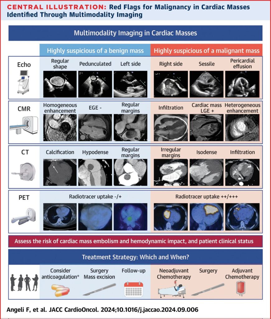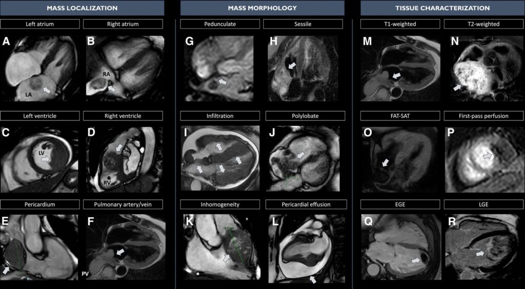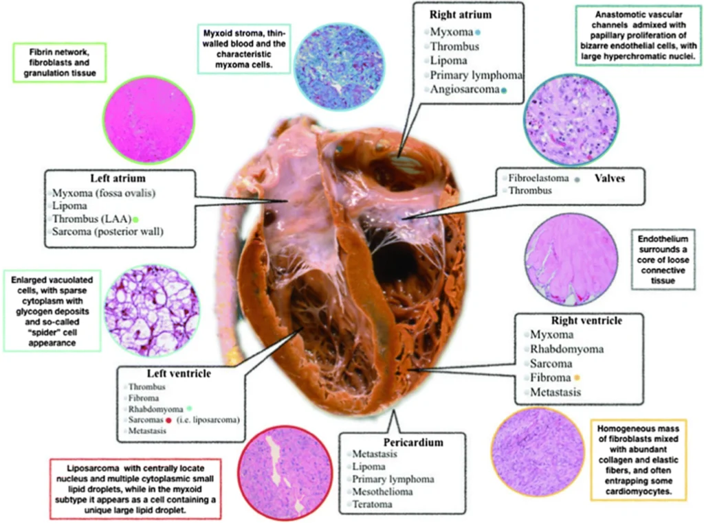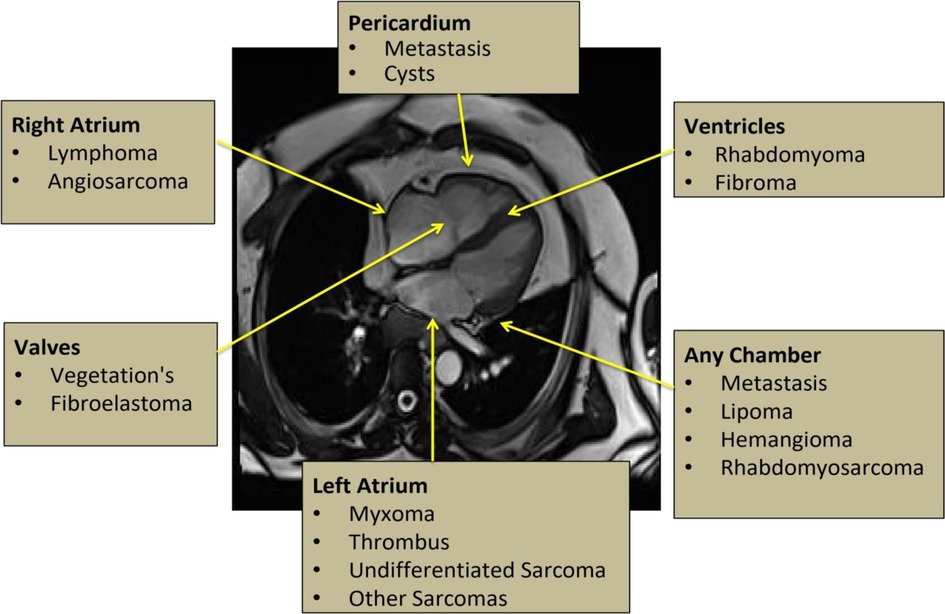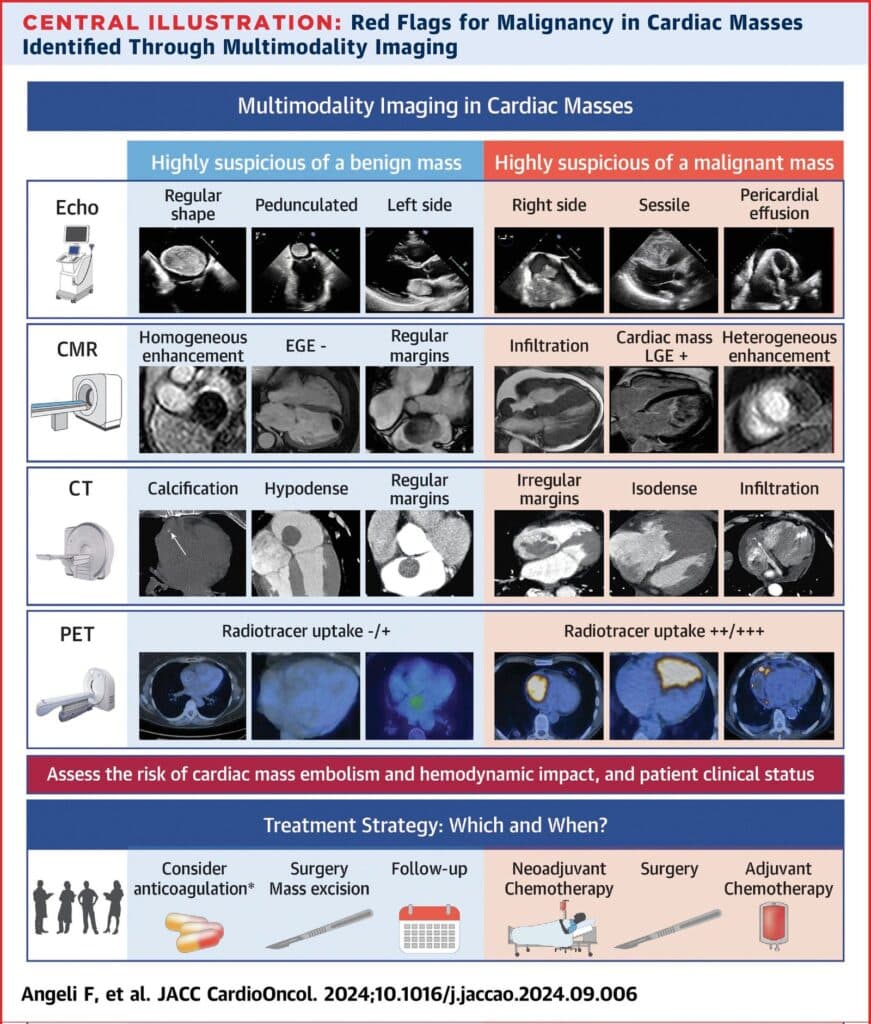
A mass on the echo report is rarely the end of the question. Thrombus, benign tumour, malignancy and normal anatomical variant can all look similar on a single modality, and the management they imply could not be more different. Multimodality imaging exists to close that gap.
What each modality contributes
- Echocardiography is almost always first: available, real-time, and good at location, attachment, mobility and haemodynamic consequence. Transoesophageal imaging adds resolution for atrial and valvular masses. Contrast helps separate avascular thrombus from a perfused tumour.
- Cardiac MRI is the reference standard for tissue characterisation. T1 and T2 weighting, first-pass perfusion and late gadolinium enhancement together distinguish thrombus, myxoma, lipoma, fibroma and malignant infiltration in a way no other single test does.
- Cardiac CT gives the best spatial resolution and the clearest view of calcification, coronary involvement and extracardiac extension — which matters when surgery is being planned.
- PET adds metabolic information: it helps separate benign from malignant, identifies the primary in suspected metastatic disease, and stages it.